Test Overview
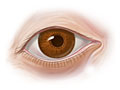
Ophthalmoscopy (also called fundoscopy) is a test that lets a doctor see inside the back of the eye, which is called the fundus. The doctor can also see other structures in the eye. The doctor uses a magnifying tool called an ophthalmoscope and a light source to see inside the eye. The test is done as part of an eye exam. It may also be done as part of a routine physical exam.
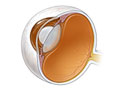
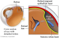
The fundus has a lining of nerve cells called the retina. The retina detects images that pass through the clear, outer covering of the eye, called the cornea. The fundus also contains blood vessels and the optic nerve.
There are two types of ophthalmoscopy.
- Direct ophthalmoscopy.
-
Your doctor uses a tool that is about the size of a small flashlight. It has many lenses that can magnify up to about 15 times.
- Indirect ophthalmoscopy.
-
Your doctor uses a small handheld lens and either a slit lamp microscope or a light attached to a headband. This test gives the doctor a wider view of the inside of the eye. It allows a better view of the fundus, even if the lens is clouded by cataracts.
Why It Is Done
Ophthalmoscopy is done to:
- Find problems or diseases of the eye, such as retina problems.
- Help find other conditions or diseases that damage the eye.
- Look for the cause of symptoms, such as headaches.
- Find other problems or diseases, such as head injuries or brain tumors.
This exam is usually done as part of a regular eye exam. Other eye tests that may be done include vision testing and testing for glaucoma.
How To Prepare
In general, there's nothing you have to do before this test, unless your doctor tells you to.
How It Is Done
Tell your doctor if you or anyone in your family has glaucoma. And tell your doctor if you are allergic to any type of eyedrops.
Before doing either type of test, your doctor may use eyedrops to widen (dilate) your pupils. This makes it easier to see the back of the eye. Your doctor may also use eyedrops to numb the surface of your eyes. It takes about 15 to 20 minutes to fully dilate the pupils.
Direct ophthalmoscopy
- You will be seated in a darkened room and will be asked to stare straight ahead at some distant spot in the room.
- Looking through the ophthalmoscope, your doctor will move very close to your face and shine a bright light into one of your eyes. Each eye is checked separately.
- Try to hold your eyes steady without blinking.
Indirect ophthalmoscopy
This type of eye exam gives a more complete view of the retina than direct ophthalmoscopy. The exam is usually done by an ophthalmologist.
- You may be asked to sit in a darkened room and to sit upright with your head on a chin rest.
- Your doctor will hold your eye open and shine a very bright light into it. The doctor will look at the eye through a special lens.
- Your doctor may ask you to look in different directions. The doctor may apply pressure to your eyeball through the skin of your eyelids by using a small, blunt tool. The pressure helps bring the edges of your fundus into view.
How long the test takes
A direct or indirect ophthalmoscopy test takes a few minutes.
How It Feels
Direct ophthalmoscopy
During this test, you may hear a clicking sound as the tool is adjusted to focus on different structures in the eye. The light is sometimes very strong, so you may see spots for a short time after the exam. Some people report seeing light spots or branching images. These are really just the outlines of the blood vessels of the retina.
Indirect ophthalmoscopy
With this test, the light is much stronger. It may be slightly painful. Pressure applied to your eyeball with the blunt tool may also hurt a little. After-images are common with this test. If the test is painful, let the doctor know.
When dilating eyedrops are used
Dilating drops may make your eyes sting and cause a medicine taste in your mouth. You'll have trouble focusing your eyes for up to 12 hours. Your distance vision usually isn't affected as much as your near vision. Your eyes may be very sensitive to light.
Risks
In some people, the dilating or numbing eyedrops can cause:
- Nausea, vomiting, dry mouth, flushing, and dizziness for a short time.
- An allergic reaction.
- A sudden increase in pressure inside the eyeball (closed-angle glaucoma).
Results
Normal: |
|
|---|---|
Abnormal: |
|
Related Information
Credits
Current as of: June 5, 2023
Author: Healthwise Staff
Clinical Review Board
All Healthwise education is reviewed by a team that includes physicians, nurses, advanced practitioners, registered dieticians, and other healthcare professionals.
Current as of: June 5, 2023
Author: Healthwise Staff
Clinical Review Board
All Healthwise education is reviewed by a team that includes physicians, nurses, advanced practitioners, registered dieticians, and other healthcare professionals.